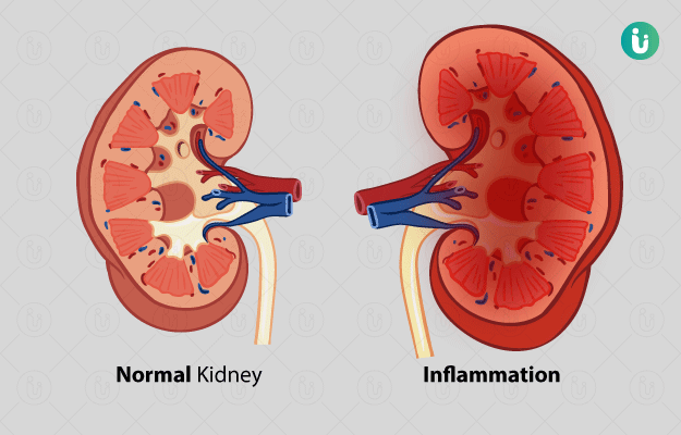
Nephritis:
- Inflammation of nephron in kidney is known as nephritis. It is inflammatory degenerative disease of kidney often referred to as ‘white-spotted kidneys’.
- Inflammation of kidney often extends to glomeruli, broadly term called as ‘glomerulonephritis’.
- It is also known as Bright’s disease.
- Glomerulonephritis can occur as a primary cause or as component of several diseases affecting several body systems such as equine infectious anemia, chronic swine fever.
- It is common causes of chronic renal failure in horses.

Classification of Nephritis:
Nephritis is classified as follows:
- Suppurative Nephritis
- Pyaemic Nephritis
- Pyelonephritis
- Non-suppurative Nephritis
- Interstitial Nephritis
- Tubular Nephritis
- Glomerulonephritis
- Specific Nephritis
- Tuberculous nephritis
Suppurative Nephritis:
- It is condition in which one or both kidneys show abscess formation.
- It is further divided into 2 types: Pyaemic nephritis (Embolic nephritis) and pyelonephritis
- Pyaemic nephritis refers to inflammation of kidney with pus formation, infection occurring via bloodstream to kidneys.
- Pyelonephritis refers to inflammation of both renal pelvis and parenchyma. Infection occurs as ascending infection from bladder to ureter and then finally to kidneys.
- Pyelonephritis is highly fatal chronic inflammation of kidneys occurring chiefly in cow but also recorded in sheep, goat, horse, buffalo and dog.
Etiology:
- Infection with pus-forming bacteria; Corneybacterium renale, Corneybacterium pyogenes, Salmonella, Shigella, Pseudomonas, Proteus
- Pyaemic nephritis- infection extending from umbilical vein, mammary gland, uterus, pericardium, lungs
- Pyelonephritis- occurs more commonly in post-parturient period, because at that time infection of uterus and vagina may occur and spread very rapidly through short and broad urethra.
Pathogenesis:
Pyaemic nephritis– Bacteria in clumps or emboli of cardiac vegetation gets disloged in glomeruli or intertubular capillaries through bloodstream where abscess is formed.
Pyelonephritis:
Infection is usually ascending in nature and associated with urinary tract infections
Obstruction of urinary tract. Due to obstruction, there is reflux of bacterial contaminated urine along ureter
Urine starts to localize in renal pelvis and then later in medulla
Bacteria produces toxin during their multiplication which causes irritation
Irritation leads to inflammation with suppuration and necrosis
Clinical Findings:
- Loss of condition over a period of week or month.
- High rise of temperature; intermittent fever
- Dullness, depression, anorexia
- Stiff gait, tenderness of lumbar region
- Palpation reveals pain in lumbar region
- Frequent micturition and straining during micturition (strangurea)
- Rough hair coat
- Rectal palpation reveals absence of lobulation in kidneys
- Hind quarter may be soiled with turbid urine
- Thick purulent, evil smelling urinary discharge
- Discharge may contain blood
- Pale mucus membrane, weak pulse
- Kidneys may be found enlarged, smooth and sensitive on rectal palpation.
Diagnosis:
- On basis of history and clinical findings
- On basis of physical examination and rectal examination
- On basis of ultrasonographic examination
- Urinalysis; high specific gravity, pus cells, W.B.C, R.B.C
Treatment:
- Broad-spectrum antibiotics should be administered. Penicillin may act well. Trimethoprimsulphadiazine may be used. Antibiotics should be used only after screening or AST.
- Diuretics; Furosemide may be used to induce polyuria @ 2-4 mg/kg, every 1-6 hours, IV/IM/SC in dogs and 0.5-2 mg/kg, b.wt. every 1-8 hours, IV/IM/SC in cats.
- Urinary acidifier or alkalizer should be used to alter pH of urine. Alteration in pH of urine helps reduce bacterial population.
- Azotemia should be treated symptomatically.
- Drugs that affects kidney such as gentamicin, OTC, neomycin are contraindicated.
Non-suppurative Nephritis:
Interstitial Nephritis:
- Accumulation of inflammatory cells in interstitial cells of kidney due to inflammation is called interstitial nephritis.
- This type of nephritis is very common in dogs, most commonly in older dogs.
- It can also be seen in swine, horses, sheep and cattle.
- It is usually found at necropsy in normal looking animals.
- It may be acute or chronic in nature.
- Interstitial inflammation without embolic nephritis or pyelonephritis is called interstitial nephritis. More recently, it is called as tubulo-interstitial nephritis.
Etiology:
- Bacteria: coli, Leptospira icterohaemorrhagiae, L. canicola
- Virus: Adenovirus, Infectious canine hepatitis virus
- Parasites: Dioctophyme renale
- Toxins: Nephrotoxic agents; lead, arsenate, sulphonamide, mercury, Amphotericin-B, paracetamol, etc.
- Autoimmunity: Tubular and interstitial lesion are being produced by formation of antitubular basement membrane antibodies
Pathogenesis:
Following infection with bacteria, bacteria gets localized in renal interstitial capillaries
They migrate through vascular endothelium, persist in interstitial spaces, migrate through intercellular junctions of tubular epithelial cells to reach renal tubular lumen.
In tubules, bacteria enter into tubular epithelial microvilli and occur in phagosomes of epithelial of PCT and DCT
These tubular epithelial cells undergo necrosis and degeneration
Clinical Findings:
- Lack of appetite
- Dull and depression
- Polyuria, polydipsia
- High temperature at initial stage
- Progressive loss of body condition
- Unpleasant breath
- Teeth, gum and tongue will be coated with reddish brown scum
- Arching of back and stiff gait
- Evidence of pain on palpation of lumbar region
- Quick, full and bounding pulse
- In chronic case, animal becomes thin and emaciated.
- Signs of dehydration, occasional vomiting
- At last, animal dies of renal failure.
Necropsy Findings:
- Kidneys have white spotted appearance- small pin-point sized grey-white circumscribed areas found scattered on cortex beneath capsule
- In diffuse nephritis, kidneys are swollen and pale with grey mottling of capsule
- Cut surface bulges
- In chronic diffuse interstitial nephritis, kidney becomes pale and shrunken
- When stripping capsule, cortex is torn.
Diagnosis:
- On basis of clinical and necropsy findings
- Urinalysis– high urine specific gravity, albumin, renal epithelial cells, WBC, RBC and casts.
Differential diagnosis:
- Disease should be differentiated with diabetes mellitus, diabetes insipidus and chronic interstitial nephritis (C.I.N)
Treatment:
- Animals should be kept at rest with minimal exercise
- Animals should be offered protein diet with high biological value
- Animals should be provided sufficient amount of water, as much as animals needs
- Dilution of urine with adequate fluid is necessary as E.coli thrives well in concentrated urine
- Course of antibiotics should be given for 7-10 days.
- Dextrose 5% with sodium chloride may be given through IV route to correct dehydration
- Parenteral B-complex should be given
- Antacid preparation should be given if gastritis results in uraemic state.
- Corticosteroid; dexamethasone given as tablet to minimize formation of immune-complex.
- Sodium bicarbonate should be given
- pH of urine should be adjusted with appropriate measures.
Glomerulonephritis:
- It denotes the inflammation of glomeruli. Inflammation of glomeruli may be followed by structural damage of other parts of kidney leading to chronic renal failure.
- Glomerulonephritis usually occurs due to immune-mediated mechanisms.
- It is characterized clinically by hematuria, proteinuria, oliguria and azotemia.
Etiology:
- Exact etiology is yet unknown but believed to be allergic, i.e. antigen-antibody reaction to foreign protein.
- It also occurs as sequale to bacterial or viral disease elsewhere in body.
- Viral infections associated with immune reactions; feline leukemia virus, feline infectious peritonitis, infectious canine hepatitis virus, equine infectious anemia in horses, bovine viral diarrhoea in cattle, swine fever (hog cholera) in pigs
- Bacterial infections such as Pyometra or pyoderma
- Chronic parasitism; dirofilariasis in dogs, Trypanosomiasis in cattle
- Auto-immune disease: canine SLE, neoplasia in dogs and cats
Pathogenesis:
Immune mediated reactions occur with formation of antigen-antibody complexes————————-> These complexes get deposited in glomerular basement membrane beneath epithelium or glomerular capillaries.————————————————————–> These complexes stimulate complement fixation reaction with formation of C3a, C5a, and C567. These are chemotactic factors for neutrophils———————————–> Neutrophils starts to gets accumulate in basement region, resulting damage of basement membrane through release of proteinase, arachidonic acid metabolites and oxygen derived free radicals and hydrogen peroxide————————————-> In later stages, monocytes infiltrate into glomeruli and causes continuing damage to glomeruli by release of biologically active molecules.
Types of Glomerulonephritis:
Acute glomerulonephritis:
- There is excessive leakage of protein in urine. It may precipitate to nephrotic syndrome.
- Affected kidneys are enlarged and pale
- Tense capsules which peels off easily
- Red dots are present on cortex indicating congested glomeruli
Membranous glomerulonephritis:
- It is most common form of immune-complex glomerulonephritis in cats.
- It is characterized by thickening of glomerular capillary basement membrane diffuse in nature.
- Affected kidneys are enlarged and pale due to fatty changes and increase in interstitial fluid caused by generalized edema
- Later, kidney becomes shrunken and fibrotic
Proliferative Glomerulonephritis:
- It is characterized by proliferation of glomerular endothelial, epithelial and mesangial cell resulting in increased cellularity of glomerular tufts
- There is accumulation of neutrophils and other leukocytes in glomeruli
Membrano-proliferative glomerulonephritis:
- It is most common form of immune-complex glomerulonephritis in dogs.
- It is characterized by hypercellularity and capillary basement membrane thickening in affected glomeruli.
- Affected kidneys are enlarged and pale
- Small red spots are visible throughout cortex.
Clinical Signs:
- Dull and depression
- Slight rise of temperature
- Polydipsia, polyuria
- Oedema (Anasarca)
- Anuria, puffy eye lids
- Hematuria
- Muscle wastage
- Proteinuria
- Vomiting
- Azotemia (Uremia)
- Dry skin coat
Diagnosis:
- On basis of history and clinical findings
- On basis of laboratory findings:
- Proteinuria is cardinal signs of glomerulonephritis
- Normocytic or normomicrochromic anemia
- Azotemia, hyperphosphatemia
- Renal biopsy
Differential Diagnosis:
- It should be differentiated from amyloidosis
- Other forms of glomerulonephritis
Treatment:
- Underlying causes should be identified and eliminated.
- Cases without nephrotic signs should be offered protein of higher biological value
- Anabolic steroid may be used to suppress immune reactions, if necessary
- Low blood volume can be corrected by administration of dextran or plasma protein
- Course of antibiotics; penicillin, ampicillin, amikacin etc. may be required against inflammatory changes
- Immunosuppressive drugs should be used. Cyclophosphamide or prednisone are used in dogs and cats.
- Regular monitoring of degrees of proteinuria, levels of urea, creatinine, and serum proteins are required.
- Antiplatelet therapy; aspirin and dipyridample are used. Aspirin @5mg/kg, b.wt. is given at 12 hours’ interval for dogs and 48-72 hours’ interval for cat.
- Diuretics can be administered to control edema and increase urinary output.