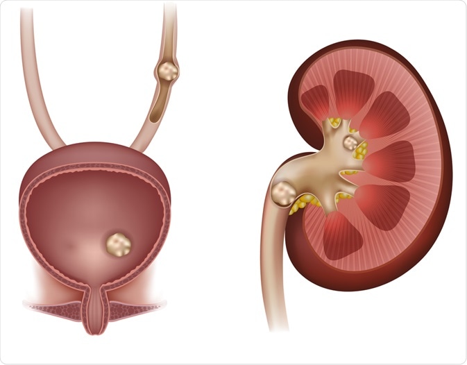
Urolithiasis:
- Deposition of crystals of various salts in any part of urinary tract is known as Urolithiasis.
- It is common as sub-clinical disorder in ruminants raised in management system where ration is composed primarily of grain, or where animals graze certain type of pasture
- Urolithiasis becomes an important clinical disease of castrated male ruminants when calculi cause urinary tract obstruction and usually obstruction of the urethra.
- Urethral obstruction is characterized clinically by complete retention of urine, frequent unsuccessful attempts to urinate, and distension of the bladder.
- Besides ruminants, it can affect variety of species including dog, cat, horse.
Etiology:
- Altered pH of urine: Low pH prevents deposition of salts, stabilizes the protective colloid and thus prevent deposition of inorganic constituents in urinary tract.
- Dehydration and inadequate supply of water. Due to inadequate water intake, urine starts to concentrate. Concentrated urine facilitate deposition of crystals in urinary tract.
- Infection of urinary tract leads to desquamation of epithelium, production of necrotic tissues, accumulation of bacteria. This along with fibrin, mucus and blood cells will form nidus or nucleus on which salts will be deposited.
- Hard water, excessive intake of calcium, Vitamin-D
- Urinary stasis and deficiency of vitamin A in diet
- Excessive intake of sulphonamides
- Ingestion or grazing on pasture species rich in oxalate, estrogens or silica.
- Metabolic disorder: Hypercalcemia, hyperparathyroidism, diets high in magnesium, phosphorus favors formation of uroliths.
Pathogenesis:
Formation of urinary stones is complex process. It involves precipitation and accumulation of minerals in urinary tract leading to stone formation. Key steps in pathogenesis of urolithiasis include:
- Supersaturation of urine:
- Supersaturation of urine occurs when concentration of certain minerals in urine exceeds their solubility limit.
- This can happen due to various factors, including dietary factors and metabolic imbalances.
- Common minerals involved in stone formation include calcium, oxalate, phosphate, and urate.
- Nucleation:
- Nucleation can occur spontaneously or be triggered by factors such as urinary stasis (a lack of urine flow) or the presence of foreign particles in the urinary tract.
- Supersaturated urine provides an environment conducive to the formation of tiny crystals (nuclei) from the excess minerals
- Crystal growth:
- Once nuclei are formed, they serve as seeds for the further growth of crystals.
- Crystals can grow over time, becoming larger and more substantial if conditions continue to favor their formation.
- Types of crystal formed depends on minerals that gets supersaturated in urine
- Aggregation:
- Crystals may aggregate or clump together, forming larger particles.
- The aggregation of crystals can be facilitated by factors such as urinary pH, the presence of mucoproteins, or cell debris in the urine.
- Matrix formation:
- In addition to minerals, urinary stones often contain organic substances, such as proteins and mucoproteins.
- These organic materials can form a matrix that binds the crystals together, contributing to stone formation and growth.
- Stone formation:
- As crystals continue to aggregate and are embedded in the matrix, they gradually form into solid stones.
- The size, composition, and location of the stones can vary, depending on the specific minerals and conditions involved in their formation.
- When stones become large enough or numerous enough, they can obstruct the urinary tract.
Occurrence of urethral obstruction:
- Obstruction may occur at any site, but most common at distal sigmoid flexure near the insertion of retractor penis muscle at steers
- In the vermiform appendage, distal to the sigmoid flexure, at the distal sigmoid flexure, or subischially in wethers or rams
- Calculi may also get lodged in urinary bladder.
Clinical Findings:
- Clinical symptoms are dependent on the location and degree of obstruction.
- Horses with complete obstruction exhibit severe abdominal pain (colic) leading to bladder or urethral rupture followed by depression and metabolic deterioration.
- Anorexia, dullness, and weight loss
- Hematuria, and stranguria
- Dysuria and incontinence
- Vomiting in dog and cat
- Severe pain on palpation
- Abdominal pain with kicking at the belly,
- Treading with the hindfeet, and swishing of the tail.
- Repeated twitching of the penis, sufficient to shake the prepuce, is often observed
- Animal may make strenuous efforts to urinate, accompanied by straining, grunting, and grating of the teeth, but these result in the passage of only a few drops of bloodstained urine
- A heavy precipitate of crystals is often visible on the preputial hairs or on the inside of the thigh
- Cattle with incomplete obstruction (“dribblers”) will pass small amounts of bloodstained urine frequently
- Rupture of the urethra or bladder occurs within 2 to 3 days if the obstruction is not relieved and the animal dies of uremia or secondary bacterial infection.
- Rupture of the urethra is more common with irregularly shaped stones that cause partial obstruction and pressure necrosis of the urethral wall.
Clinical Signs in Rams with obstruction of urethra:
- Sudden depression
- Inappetence
- Stamping the feet
- Tail swishing
- Kicking at the abdomen
- Bruxism
- Anuria or the passage of only a few drops of urine are common.
Diagnosis:
- Based on history of diet or grazing
- Based on clinical finding
- Based on physical examination of urethra, palpation of bladder
- Urinalysis: Detection of crystal on examination of urine
- Laboratory findings: Increased SUN and creatinine concentrations, hyponatremia, hypochloremia, hyperphosphatemia
- Ultrasonograhic examination of bladder
- Radiographic examination of urinary tract
Treatment:
- The treatment of obstructive urolithiasis has traditionally been primarily surgical, including urethral process amputation (rams, wethers, bucks, llamas, and alpacas), prepubic and perineal urethrostomy, laser lithotripsy, tube cystotomy, and bladder marsupialization.
- Rams, bucks, and wethers should all have their glans penis exteriorized and inspected and the urethral process amputated using a scalpel blade. It is best accompanied by having an assistant restrain the animal in sitting position. Penis is exteriorized by grasping shaft of penis within prepuce and retracting prepuce to expose tip of penis which is then grasped with a gauze sponge.
- Alteration of pH of urine helps to dissolve uroliths within bladder. Instillation of 30-200 ml of acetic acid or hemiacidrin solution through a cystotomy catheter or long needle into the bladder after removal of most but not all the urine in the bladder have resolved problem
- In case of infection, suitable antibiotics based on culture reports should be administered.
- Blood urea in small animal are controlled with ciploric or Zyloric tablet @ 100mg, BID, orally
- Cystone tablet @ 2 tablet may be tried.
- Complete obstruction requires surgical intervention. Perineal urethrostomy is done to remove calculi and relieve pressure on bladder
Prevention:
- Prevention is essential in managing urolithiasis. Diet modifications, supply of clean and free drinking water, regular checkup helps in identifying problems.
- The diet should contain an adequate balance of calcium and phosphorus to avoid precipitation of excess phosphorus in the urine.
- Water intake can be promoted by supplementing the ration with salt.
- The feeding of ammonium chloride at 0.5%–2.0% of dry matter intake, approximately 45 g/day to steers, 10 g daily to sheep, and 0.4–0.5 g/kg BW each day to male goats) may prevent urolithiasis caused by struvite or calcium carbonate