Types of Acute Inflammation:
Based on the main component of exudate, acute inflammation are classified as follows:
- Catarrhal or Mucous Inflammation:
- Main component of exudate is mucus.
- Occurs in those areas where cells are capable of producing mucin.
- Produced by irritants of mild nature, mildly irritating chemicals, irritating foods in digestive tract, inhaled dust, cold air.
- There is no proliferation of epithelium which is desquamated into exudate. These exudate contains desquamated cells, neutrophils, and mucus.
- Mucus are transparent, clear, and glistening slimy material containing water and mucin.
- If cause is removed, recovery occurs quickly.

- Serous Inflammation:
- Main component of exudate is plasma (derived from blood) or clear watery fluid derived from secretions of mesothelial cells lining peritoneal, pleural, and pericardial cavities.
- Irritants are moderately severe.
- Second-degree burns, traumatic injuries of rubbing nature are examples of serous inflammation where there is blister formation.
- It is first stage in main inflammatory processes.
Outcome is favourable if cause is removed. If it persist, exudate usually gets organized and adhesion may form

Fig: Serous inflammation of larynx
3. Fibrinous Inflammation:
- Main component of exudate is fibrin.
- It is caused by violent type of injury. Due to increased vascular permeability, fibrinogen escape to surrounding tissues.
- Seen in viral disease like infectious feline enteritis and malignant catarrhal fever and when mucous membranes are invaded by Corneybacterium diptheriae, Salmonella.
- Organ is firmer and tenser than normal.
- Especially seen in lungs in case of pneumonia. Fibrin gets accumulated in alveoli of lungs, due to which lungs acquires both consistency and appearance of liver.
- Fibrin appears as stringy, yellowish, net-like material on epithelial surface.
- Occurs commonly in body cavities such as pleural and peritoneal sac.
- Masses of fibrin on epithelial surface may either form pseudomembrane or diphtheritic membrane.
- As there is extensive tissue destruction, animal may not survive.
- Exudates may be degraded by fibrinolysis and accumulated debris may be removed by macrophages, resulting in restoration of normal tissue structure (resolution). However, extensive fibrin-rich exudate may not be completely removed and replaced by ingrowth of fibroblast and blood vessels (organization), leading ultimately to scarring.

4. Suppurative inflammation:
- Characterized by production of pus or purulent exudate which results from softening and liquefaction of tissues.
- This is seen in bacterial infection of pyogenic nature.
- Pus is composed of necrotic neutrophils, necrotic tissue cells, inflammatory exudate including serum.
- Pus are alkaline and usually creamy in colour. They may be:
- White or yellow: streptococcal and staphylococcal infection
- Greenish: Corneybacterium in cattle
- Blue-green: Pigment forming pyocyaneus bacillus
- Black: From disintegrating hoof material.
- Lysosomal enzymes are present in pus and extent of proteolysis they produce determines the viscosity of pus.
- Canine pus- Thin watery- Due to extremely proteolytic neutrophilic enzyme
- Bovine pus- Viscid
Avian pus- Dry, caseous- Due to presence of antienzymes
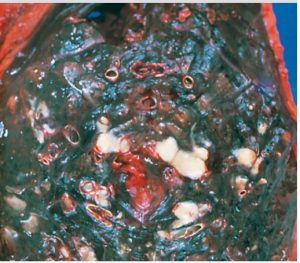
Fig: Suppurative Inflammation of Bronchitis
5. Haemorrhagic inflammation:
- Main component of exudate is erythrocyte.
- Caused by violent type of irritant.
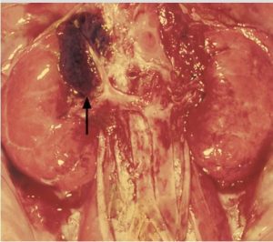
Fig: Haemorrhagic Inflammation of Adrenal Cortex
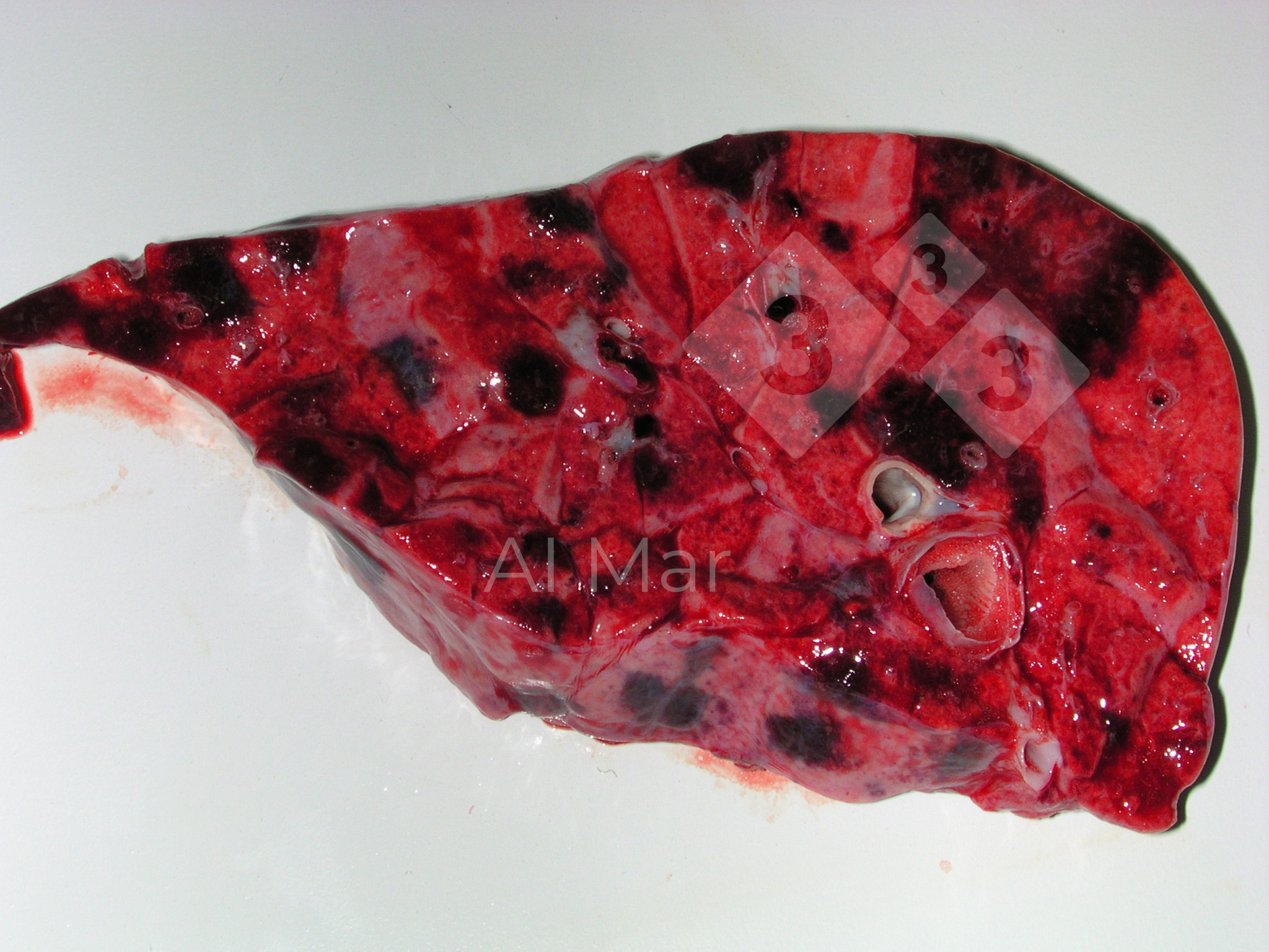
Fig: Haemorrhagic Pneumonia