Neoplasm :
Introduction :
- Neoplasia = new growth ( abnormal and excessive growth )
- Study of neoplasm (tumor) = oncology
- Neoplasm is the new and abnormal growth of tissue in a part of the body , especially as a characteristic of cancer.
- It is a self controlling growth formed by unlimited multiplication of abnormal cells.

Classification :
The classification is done under following basis :
a. Behaviour of tumor ( benign or malignant )
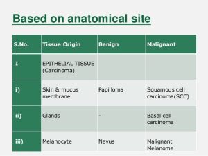
i. Benign : epithelial
- Mesenchymal
ii. Malignant : primary
- Epithelial ( carcinoma )
- Mesenchymal ( sarcoma)
b. On the based on anatomical site
i. Histological analysis ( grading )
ii. Extent of disease : classification ( staging )
iii. TNM ( tumor , node, metastasis )
Behaviour of tumor
A. Benign tumor : ( pathology)
I. Gross finding :
- Size : usually small in size
- Shape : usually ovoid or rounded in shape
- Capsule : usually encapsulated
- Cut section solid or cystic
- Haemorrhages and necrosis are usually absent .
II. Microscopic:
- Differentiation : the cells are well differentiated i.e. tumor cells closely similar to tissue of origin .
- Nucleocytoplasmic ratio ( N/C ) : small / normal
- Stroma : is usually well formed with few blood vessels.
III. Behaviour :
- Rate of growth : usually slow
- Mode of growth : expansion
- Localization : usually localized
- Effects on host : usually do not destroy surrounding structures and don’t kill the patient (except in certain sites as in brain )
- Recurrence : usually not recurrent
- Metastasis : don’t metastasis ( development of secondary growth at a distance from a primary site of tomor )
- Malignant change : may occur
Nomenclature :
- Benign tumor : prefix + suffix
Type of cell + ( oma )
- Benign tumor arising in fibrous tissue = fibroma
- Benign tumor arising in fatty tissue = lipoma
- Benign epithelial tumors : Papilloma
Adenoma ( benign epithelial neoplasms producing gland pattern )
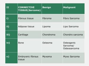
- Mesenchymal tumors :
- CT tumors :
- Fibroma
- Lipoma
- Chondroma
- Osteoma
- Muscle tumors :
- Leiomyoma ( also known as fibroids, is a benign smooth muscle tumor that very rarely becomes cancer (0.1%))
- Rhabdomyoma ( is a benign tumor of striated muscle)
- Vessels :
- Haemangioma ( is a usually benign vascular tumor derived from blood vessel cell types.)
- Lymphangioma ( are malformations of the lymphatic system characterized by lesions that are thin-walled cysts; these cysts can be macroscopic, as in a cystic hygroma)
B. Malignant tumors :
- Have two basic component :
- Parenchyma :
- Made up of neoplastic cells
- Determines the biological behaviour of the tumor from which the tumor derives its name .
- Stroma :
- Made up of non-neoplastic cells, host derived CT and blood vessels
Malignant tumor arising in mesenchymal tissue ( multipotent stem cells found in bone marrow that are important for making and repairing skeletal tissues, such as cartilage, bone and the fat found in bone marrow.)
Sarcoma
- Fibrous tissue : Fibrosarcoma
- Bone : Osteosarcoma
- Cartilage : chondrosarcoma
Malignant tissue arising from epithelial origin : Carcinoma
- Squamous cell carcinoma
- Renal cell adenocarcinoma
- Cholangiocarcinoma ( bile duct cancer )
Nomenclature of some malignant tumors with exception :
- Melanoma ( skin)
- Mesothelioma ( mesothelium )
- Seminoma ( testis)
- Lymphoma ( lymphoid tissue )
Pathology :
a. Gross lesions :
- Size : usually reach large size
- Shape :
- Polypoid of fungating mass in tumors of solid organs .
- Malignant ulcer in tumor of surface epithelium
- Infiltrating annular mass in tumor of hollow organs
- Capsule : non- capsul;ated
- Cut section solid / cystic
- Haemorrhage and necrosis : common
b. Microscopic pathology :
- Differentiation : the cells dhow loss of differentiation
- The cells show some or all features of mal;ignancy as loss of polarity of cells, hyperchromatic nuclei , increase N/C ratio , abnormal mitosis and prominent nucleolus
- Stroma : usually desmoplastic with rich vascularity
Behaviour :
- Rate of growth : usually rapid
- Mode of growth : by infiltration
- Localization : usually not localized
- Effect on host : can kill the patient whenever present
- Recurrence : may occur
- Metastasis : may occur
Precancerous lesions :
- Chronic inflammatory lesions
- Hyperplastic lesions
- Some benign tumors
- Other lesions as peptic ulcer and undescribed testis.
Spread of malignant tumors :
a. Mechanism of spread
- Invasion of matrix
- Vascular dissemination and homing of tumor cells
b. Route of spread :
i. Direct / local spread
Malignant cells infiltrate the surrounding structures in all direction
ii. Distant spread :
- Lymphatic spread
- Lymphatic permeation
- Lymphatic embolization
- Blood spread
- Course of tumor emboli
- Organ metastasis
- Transcoelomic spread
- Spread by implantation
- Anatomical; site :
- Histological analytic :
It defines the types of tissue from which tumor is originated :
- Grade I : cells are slightly differed from normal cells and well differentiated
- Grade II : cells are more abnormal and moderately differentiated.
- Grade III : cells are abnormal and poorly differentiated
- Grade IV : cells are immediate and primitive and undifferentiated
Staging : progression or spread in body
Grading : cell differentiation and rate of growth – microscopy
- Extent of disease :
- Stage 0 : cancer in situ (cell)
- Stage I : tumor linked to tissue origin , localized tumor growth
- Stage II : limited local spread
- Stage III : extensive local and regional spread
- Stage IV : metastasis
- TNM classification :
- This system represent clinical staging of cancer
- Used to determine the extent of disease process of cancers according to three parameters:
- Tumor size ( T)
- Degree of regional spread to LN ( N)
- Presence of metastasis (M)
- It has been used in diagnosing breast cancer .
Etiology :
A. Benign tumors :
- Often the cause is unknown
- But the growth might be linked to :
- Environmental toxins such as exposure to radiation
- Genetics
- Diet
- Stress
- Local trauma or injury
- Inflammation or infection
B. Malignant tumors :
a. Precancerous lesions :
Some lesions that exhibit a tendency to undergo malignant transformation
- Endometrial hyperplasia – endometrial carcinoma
- Liver cirrhosis – hepatocellular carcinoma
- Squamous metaplasia lead to squamous cell carcinoma
- Benign tumors :
- Papilloma of urinary bladder
- Adenoma of thyroid or colon
b. Helping factors ( carcinogens)
- Age
- Sex
- Diet : fat – colonic cancer
- Smoked fish – gastric carcinoma
- Smoke : lung cancer
- Heredity : retinoblastoma
c. Carcinogens :
- Chemical carcinogens
- Viruses
- Radiation
Mode of growth :
a. Benign tumors :
- Remain localized
- Grow by expansion with available space or by compression of tissues
- Cause adverse effects if near vital structure
b. Malignant tumors :
- Do not remain localised
- Local growth is by infiltration and super population of adjacent of tissues
- Invasion causes destruction of local normal tissues.
Development / pathophysiology :
Carcinogens are substances that when introduced into cells cause changes in structure and function of cells and lead to cancer , however it occurs in several stages over a period of time .
- Three identified status :
- Initiation
- Promotion
- Progression
- Initiation
Causative agents ( carcinogens)
⬇
Target cell
⬇
Altered
⬇
Cancer
- Promotion
⬇
Proliferation at mitotic rate at tissue of organ
- Progression
⬇
Evidence of clinical disease
⬇
Evidence of regional spread and metastasis
Diagnosis :
- Morphological methods :
- History
- Clinical sign
- Biochemical assay : useful for measuring the levels of tumor associated with enzymes , hormones , tumors markers in serum
- Biopsy
- Radiograph
- Molecular diagnosis : PCR , FISH ( Fluorescent in situ Hybridization )