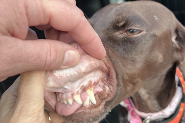
Anemia:
- It may be defined as decrease in quantity of hemoglobin or number of erythrocytes or both per unit volume of blood.
- It is characterized clinically by pale mucus membrane, tachycardia and loss of muscular strength and vigour.

Classification of anemia:
Anemia are classified based on following categories;
- According to morphology
- According to response
- According to etiology/patho-physiologic mechanism
Anemia according to morphology of red blood cells:
On the basis of morphology of red blood cells, anemia are of following types:
- Macrocytic normochromic anemia:
- This type of anemia denotes the presence of immature red blood cells in blood.
- It occurs due to deficiency of vitamin B12, folic acid, cobalt, intrinsic factors and erythrocyte maturation factors.
- Size of RBCs is increased in this type of anemia and bone marrow is responsive.
- Macrocytic hypochromic anemia:
- This anemia occurs in regenerative phases after hemorrhage.
- Hemorrhage may be due to trauma, wound, surgical bleeding, parasitic oozing of blood, epistaxis, etc.
- Size of RBC is increased with decreased Hb concentration.
- Normocytic normochromic anemia:
- This is known as aplastic or hypoplastic anemia.
- Staining properties of RBC’s are normal. Number of granulocytes and thrombocytes are reduced
- It occurs due to suppression of bone marrow activity in acute or sub-acute systemic disease.
- It may be primary or secondary. Primary is rare.
- Secondary anemia may occur due to:
- Chronic hemorrhage
- Neoplasm
- Deficiency of vitamin-6 and prothrombin
- Ionization and irradiation
- Chemical poisoning
- Bracken fern poisoning
- Sulphonamide and chloramphenicol toxicity
- Size of RBC is normal with normal hemoglobin concentration.
- Normocytic hypochromic anemia:
- This type of anemia occurs due to reduced hemoglobin formation.
- Size of R.B.C is normal with reduced hemoglobin concentration.
- This anemia occurs due to following factors/causes:
- Dietary deficiency of iron; due to exclusive intake of milk in piglets or defective absorption of iron
- Dietary deficiency of copper
- Dietary deficiency of ascorbic acid (Vitamin-C)
- Dietary deficiency of pyridoxine
- Dietary deficiency of nicotinic acid
- Dietary deficiency of riboflavin
- Deficiency of thyroxine
- Microcytic normochromic anemia:
- In this type of anemia, size of RBC is reduced with normal hemoglobin concentration
- This anemia occurs due to following causes:
- Chemical poisoning
- Chronic interstitial nephritis
- Worm infection
- Chronic infection like tuberculosis, brucellosis
- Ionizing radiation
- Microcytic hypochromic anemia:
- This anemia result due to deficiency of iron.
- It also occurs due to dietary deficiency of copper, manganese, cobalt, ascorbic acid, pyridoxine, nicotinic acid, riboflavin, thyroxin, etc.
- Size of RBCs are reduced with reduced hemoglobin concentration.
Anemia according to response:
- Regenerative anemia:
- Bone marrow is responsive to this type of anemia.
- It is characterized by increased number of immature RBC in peripheral circulation
- Ex; blood loss anemia, hemolytic anemia
- Non-regenerative anemia:
- Bone marrow is not responsive to anemic state.
- Bone marrow are unable to produce red blood cells.
- Ex; Depression anemias and aplastic anemias
Anemia according to etiology and pathophysiologic condition:
- Haemorrhagic anemia:
- There is loss of RBCs in this type of anemia. Blood loss exceeds production.
- It occurs when 25-40% of blood is lost. This condition leads to hypochromic, microcytic anemia
- It may be acute or chronic in nature
- Acute haemorrhagic anemia occurs due to:
- Any wound
- Epistaxis
- Hemoptysis
- Surgical bleeding
- Splenomegaly in dog
- Brackern fern poisoning
- Sweet clover poisoning
- Rapid X-ray exposure
- Gastrointestinal hemorrhage
- Genito-urinary hemorrhage
- Chronic hemorrhagic anemia occurs due to:
- Heavy ectoparasitic infestation
- Endoparasitic infection
- Deficiency of vit. K, vit.C and prothrombin
- Coccidiosis in young animals
- Enzootic bovine Hematuria
- Hemolytic anemia:
- This type of anemia occurs due to accelerated erythrocyte destruction.
- It is characterized by macrocytic hypochromic anemia.
- It may be of 2 types; congenital hemolytic anemia and acquired hemolytic anemia
- Congenital anemia occurs due to defect in formation of stroma of protein or hemoglobin. It is heritable disease due to simple Mendelian recessive gene.
- Acquired hemolytic anemia occurs due to:
- Viral infection: Equine infectious anemia, infectious mononucleosis
- Bacterial infection: Leptospirosis, Clostridium haemolyticum, Clostridium perfringens type A, streptococcal and staphylococcal infection
- Protozoal infection: Babesiosis in all species, Anaplasmosis in ruminants, theileriosis, haemobartonellosis in dog and cat
- Phosphorus deficiency
- Copper poisoning
- Phenothiazine toxicity
- Ingestion of arsenic, bismuth, lead
- Excessive use of sulphonamides and other aspirin drug
- Snake venom
- Immunological haemolytic anemia:
- It is of 2 types; Autoimmune haemolytic anaemia and iso-immune haemolytic anemia
- Auto-immune hemolytic anemia:
- It is recorded in dog and cat
- Under special circumstances, there may be mutation of genes and protein of own body becomes antigen and body produced antibody against own erythrocytes.
- Due to result of Ag-Ab reaction, hemolysis occurs
- Disease is characterized by sudden onset of anemia and spherocytosis
- Iso-immune haemolytic anemia:
- This anemia occurs due to transfer of maternal iso-antibodies from dam to new-born through colostrum.
- Dyshaemopoetic anemia:
- This anemia results due to depression of erythropoiesis
- Certain chronic suppurative process may depress bone marrow activity leading to less production of erythrocytes.
- It may be observed in case of nephritis, chronic interstitial nephritis, neoplastic disease, rapid exposure to X-ray or other radioactive substances, drug toxicity, viral infection causing marrow suppression.
Clinical Signs:
- Paleness of visible mucus membrane
- Muscular weakness, dull and depression
- Inappetance to anorexia
- Tachycardia (increased heart rate)
- Decrease in intensity of heart sound in later stage
- Labored breathing at terminal stage due to anemic anoxia
- Signs of shock in hemorrhagic anemia
- Jaundice
- Hemoglobinuria, hematuria, edema
Laboratory findings:
- Reduction in number of RBC
- Reduction in amount of hemoglobin
- Morphological abnormalities in RBC
Diagnosis:
- Based on history, clinical findings and laboratory findings
Treatment:
- Attempt should be made to correct primary causes with appropriate measures
- In severe cases, whole blood transfusion is carried out.
- Plasma extender; haemacel may be used @ 10-15ml/kg, b.wt.
- Haematinic preparations may be used in less severe cases and as supportive treatment after transfusion.
- Iron preparation; ferrous sulphate are injected IV, gives rapid response. Ferrous sulphate, 4-8 g, 1 dose BID for 3 days
- Fersolate tablet @ 1-2 tab, BID for dog
- Imferon injection available in 2 ml ampule. Sig; 4 amps (6-8ml) at a time daily or alternate days according to severity through IM route
- Iron dextran @ 5-10 ml. I/M
- Liver extract; Belamyl injection, Livogen injection @ 5-10 ml in alternate days in case of severe anemia.
- Livogen syrup @ 1-2 t.s.f, BID, PO for small animals
- In case of immune mediated hemolytic anemia, glucocorticoid therapy is given. Prednisolone @ 1-2 mg/kg, PO, BID.
- Dexamethasone is given @0.3-0.5 mg/kg, IV, OD
- For immunosuppression in dogs and cats, azathioprine is used @ 2mg/kg, PO, OD. Cyclosporine can be used @ 5-10 mg/kg, b.wt., PO, BID