Balantidium coli
Location and host
- This species occurs world-wide in caecum and colon of pig, man, chimpanzee, monkey and other non-human primates and dog. The species causes balantidial dysentery in man and other host.
- Most commonly occurs in pigs.
Morphology
- One of the largest protozoan parasites parasitizing man.
- Two stages: Trophozoite stages (vegetative form) and cystic stage.
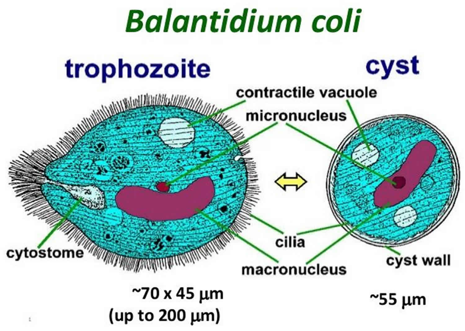
a. Trophozoite (vegetative form):
- Largest form and average size of 50-60 µm in length but sometimes, they reach upto 150 µm in length and easily observed in the stool of infected animals.
- It is ovoid, egg-shaped, pear-shaped or occasionally subcylindrical.
- Body surface is covered with slightly oblique longitudinal rows of cilia.
- Peristome is sub-terminal at narrower end which leads to a tiny mouth called cystotome. Posterior end has excretory organelle called cytopyge.
- It has two nuclei: Macronucleus and micronucleus. Macronucleus is large kidney shaped or sausage-shaped. Micronucleus is small and lies in depression of macronucleus and rounded shaped.
- Two contractile vacuoles, one being terminal and other near the center of the body. The food vacuoles are numerous containing starch grains, cell fragments, bacteria and erythrocytes.
- Organism is actively motile and moves quickly when examined under the microscope.
- Under conditions like constipation and bowel dehydration, organism undergo encystment inside the host. They may also undergo encystment outside the host body.
b. Cyst stage:
- Ovoid to spherical and measure 40-60 µm in diameter.
- Light yellowish or greenish in color, with hyaline cytoplasm
- Cyst wall has two membranes.

Fig: Trophozoite of B. coli

Fig: Cyst stage
Life cycle
- Infection of host occurs through ingestion of cystic stage
- After reaching intestine, excystation of cyst takes place.
- Excystation produces trophozoite from cyst
- Parasite multiplies by transverse binary fission but conjugation also occurs.
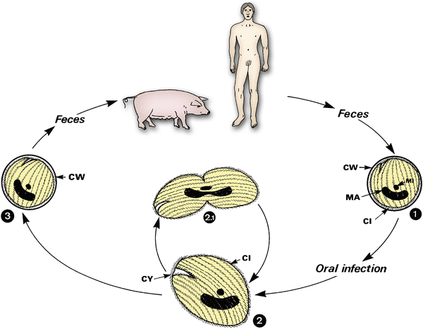
Mode of transmission
- Ingestion of contaminated food and water.
Pathogenesis/ Clinical signs
- Parasite live as commensal in lumen of large intestine and don’t penetrate intact intestinal mucosa. In case of heavy infection, it may invade the mucosa and cause superficial and even deep ulcerations. It causes mild to severe enteritis.
- In acute infection, it causes hemorrhagic dysentery.
- In human being, it is pathogenic and causes diarrhea and dysentery.
- Ulcers in intestine are infiltered with round cells and areas of coagulative necrosis and hemorrhagic area may be present.
- Dogs also suffer severely from balantidial infection. There may be diarrhea or dysentery for several days.
Diagnosis
- Detection of cysts in faeces.
- Histological examination of intestinal lesion.
Treatment / Chemoprophylaxis
- Oxytetracycline @0.5 gm orally
- Carbasone @ 250 mg daily for 10 days
- Diodoquin 300 mg once daily for 5 days