Marek’s Disease:
Syn: Range paralysis, Skin leukosis, Fowl paralysis, Polyneuritis
- It is highly contagious, lympho-proliferative disease of poultry.
- It is characterized by marked enlargement of nerves, liver, spleen and kidneys due to diffuse growth of certain cells (lymphoid cells).
Etiology:
- Alphaherpesvirus of Herpesviridae family
- Virus is enveloped, ds DNA, hexagonal in shape, size; 85-100µm
- 3 serotypes are recognized: serotype 1, 2 and 3
- Serotype-1; disease producing strain, serotype-2: non-disease producing strains, serotype-3: non-disease producing strain related to turkey
- Serotype-1 further classified as:
- Mildly harmful (MMDV)
- Harmful (vMDV)
- Very harmful (vvMDV)
- Very very harmful (vv+ MDV)
- It is strictly cell-associated virus, except in feather follicles, where cell-free virus is produced.
Epidemiology:
- Disease was first described by Hungarian Veterinarian Jozsef Marek in 1907.
- Outbreaks were reported in 1914 in USA, Netherlands, Great Britian and many other countries.
- It is distributed worldwide and has got economic importance.
- It usually occurs between 2-8 months of age; however, it has also been reported in egg producing birds.
- In 22 outbreaks in Haryana, average mortality was 32.4% and seroprevalence of 78.4%.
- It was a major threat to commercial poultry of Nepal during 1998-2002; 70% mortality and 40% drop in egg production was seen.
- Major outbreaks were reported from Chitwan, Nawalparasi, Kathmandu, Lalitpur, Bhaktapur, Kavre, Rupandehi, Syangja, Kaski and Nuwakot.
Transmission:
- Inhalation of infected material from the environment.
- Virus particle can persist for a considerable period of time in dandruff of feather follicles.
- Virus is also present in oral, nasal and tracheal secretions
- Darkling beetle (Alphitribus diaperinus) can carry the virus for several weeks.
- Virus is not transmitted through egg.
Pathogenesis:
- Chickens of 12-14 weeks of age are most susceptible to Marek’s disease.
- Generally, it doesnot occur in chickens below 6 weeks of age and older birds above 24 weeks of age.
- 3 phases of viral infection are present:
- Productive-restrictive infection
- Latent infection
- Neoplastic transformation
- Virus enters through the respiratory tract, by inhalation.
- Virus grows within certain cells of the lungs.
- It is followed by cytolytic infection of lymphoid system, mostly in Bursa and Thymus. Lymphocytes are destructed mainly in Bursa of Fabricius, thymus and also spleen.
- After 6-7 days, infection becomes latent. Cell-mediated immunity has been shown to be important. Most latently infected cells are CD4+ T-cells, although CD-8+Tcells and B-cells.
- Virus is spread throughout body by infected lymphocytes and present in blood in a cell-associated form.
- Secondary destructive infection occurs in feather follicles 2 weeks after primary infection and cell-free virus is produced and shed into environment through feather debris and dander.
- Proliferation of lymphocytes is final response and progress to tumor formation. T-lymphocytes transform into tumor cells and proliferate in nerves and other tissues and organ.
Clinical Findings:
Disease appear in several forms:
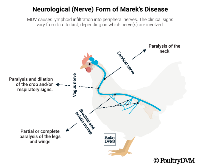
a. Classical form or Neural form:
- Mostly noted at onset of sexual maturity, i.e. about 16 weeks and at time of peak laying, i.e. 30 weeks of age.
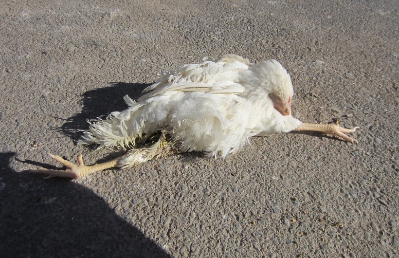
- Paralysis of legs, drooping of wings
- Birds are unable to stand.
- Legs and wings are stretched in either direction.
- Split leg stance is usual feature.
- If cervical nerves are affected, there may be torticollis.
b. Acute or Visceral form:
- Depression, droopiness, unthriftiness, dehydration
- Emaciation, anemia
- Mortality rate may go as high as 60%
- Chicks may die on all of sudden without any signs.

c. Transitional Paralytic form:
- It occurs in chicken at age of 5-18 weeks of age.
- Sudden development of paresis
- Paralysis of legs, wings and neck
- Sign usually disappear within 24-28 hours.
d. Ocular form:
- Blindness develops in the birds.
- Grey eye or pearl eye due to cell infiltration.

e. Skin or cutaneous form:
- Distinct white nodules are found on the skin.
- In extreme cases, it looks as brownish scales.

f. Muscular form:
- Muscles look lustreless, whitish grey.
- Tiny streaks to nodular tumors in skin.

Post-Mortem Findings:
- Marked enlargement of one or more nerves. Most commonly sciatic and brachial plexus; 2-3 times normal thickness.
- Striation and glistening appearance of nerve is lost.
- Nerve may appear greyish and sometimes edematous.
- Cauliflower like appearance of ovary due to growth of tumor.
- Lymphomas are sometimes seen in liver, heart, kidney and lungs.
- Marked enlargement of liver and spleen.
- Significant enlargement of proventriculus.
- Heart is pale, have single or nodular tumors in myocardium.
- Atherosclerosis, lipid accumulation in arteries.
- Degeneration of bone marrow.

Diagnosis:
- Based on clinical findings and PM findings
- Isolation of virus in chick embryo inoculation
- Demonstration of tumor-associated surface antigen (MATSA) on cells by immunofluorescence.
- Virus neutralization test (VNT), FAT, IFAT
- Immunoperoxidase test
Differential Diagnosis:
- In lymphoid leukosis, chickens usually have gross tumors in Bursa of Fabricius. Tumors are absent in Marek’s disease.
Treatment:
- There is no effective treatment available.
- Symptomatic treatment for paralysis may be adopted.
Control Measures:
- Isolation of growing chickens from source of infection.
- Farm should be disinfected with formalin and house should be kept vacant for about a month.
- All in and all out method of rearing should be adopted.
- Insecticides should be used to prevent insect load in the farm since virus may be transmitted through insects.
- Attempt should be made to evolve a MD resistant stock by careful breeding and repeated testing.
- Vaccination of flock; 3 types of vaccines are used; attenuated MDV, avirulent MDV and turkey herpesvirus (HVT). HVT is widely used.
- Living, HVT, FC-126 strain; 0.2 ml SC in back of neck in day-one old chick.