Tetanus:
Syn: Locked Jaw, Saw-horse stance
- It is an acute, non-febrile, infectious disease of mammals caused by bacterial toxins.
- It is characterized by spasmodic contraction of skeletal muscles, death due to respiratory arrest or convulsion.
Etiology:
- Clostridium tetani
- Organism is long, slender, rod shaped anaerobe with rounded ends
- Size; 2×0.5 µm
- Organism develops terminal spores which gives it drumstick appearance.
- Gram+ve, motile, spore forming bacteria
- Spores are resistant to heat, drying and remain in soil for years.
- Spores can be destroyed by boiling at 115°C for 30-60 minutes.

Epidemiology:
- Organism is more common in tropics than in temperate zones.
- It is sporadic disease in Nepal, India
- Organism produce three types of toxin; tetanospasmin (neurotoxin), tetanolysin (haemolysin) and fibrinolysin.
- Horses and mules are most susceptible to tetanus followed by sheep, goat, pigs. Cattle are less susceptible.
- Birds are resistant to tetanus due to their high body temperature, high metabolic rate.
- Morbidity is very low 1-2% while mortality is high; 80% if left untreated in affected animals.
Transmission:
- Through contaminated soil, animal manure
- Organism gains entry through deep punctured wound
- During parturition, manual handling of genitalia with contaminated hand
- Through ROP and prolapse if exposed to contaminated soil
- Through castration by open methods, shearing, docking
- Through contaminated umbilicus in case of neonates
Pathogenesis:


Clinical Signs:
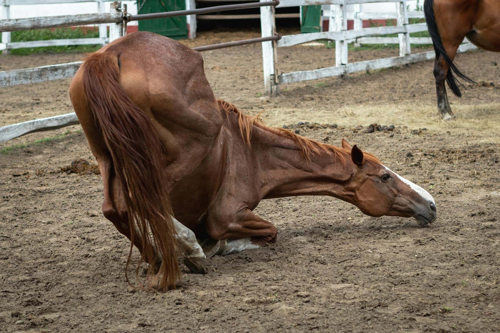
Horse:
- Stiff gait and apathy to feed is initial sign of the disease.
- Characteristic “saw-horse stance” develops.
- Prolapse of third eyelid
- Erection of ears, immobility of ears
- Horse stands with its feet widely apart and its head and neck in an extension position.
- Rigidity of facial muscles gives an anxious appearance.
- Spasm of masseter muscle leads to trismus (lock jaw).
- Mouth is held tightly closed and separation of jaw is hardly possible.
- There is restriction of mastication and dribbling of saliva from mouth.
- Animal remains hypersensitive. Light noise, air current, aggravate the spasm of muscles.
- Violent trembling may occur sometimes due to hypersensitivity.
- Profuse sweating all over the body
- In acute case, temperature and pulse rate may be elevated but sub-acute case will show normal level.
- Finally, horse is unable to stand and dies due to asphyxia within 3-10 days.
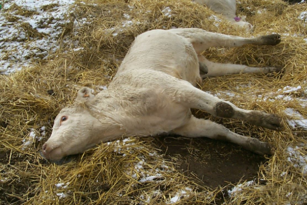
Cattle and Buffalo:
- Initial signs include restricted movement, muscular stiffness and difficulty in walk.
- Fall in milk yield
- Lock jaw and hypersensitivity on little stimuli
- Prolapse of third eyelid
- Pump handle position of tail
- Finally, lateral recumbency supervenes along with extensor rigidity and opisthotonus
- Rumen may appear like drum.
- Cow dies due to asphyxiation.

Sheep and goat:
- Dullness
- Head drawn on one side or backward
- Opisthotonus
- Hypersensitivity to noise
- Tonic contraction of masseter muscle.
- Death occurs due to asphyxiation.
PM Findings:
- Lesion is not characteristic.
- There is setting up of early rigor mortis.
- Congestion in spinal cord, medulla and nerves.
- Lungs remain hyperemic.
- Hemorrhages in skeletal muscles, loss of cross striations and atrophy of myofibrils.
Diagnosis:
- Clinical signs are so suggestive, it is hardly difficult to diagnose.
- Identification of bacteria through gram staining.
- Inoculation of mice with cultured materials from wounds and produce signs of tetanus in unprotected mice.
- In lab, organism may can be cultivated by anaerobic method in meat broth or blood agar.
- Serum antitoxin antibody levels can be measured.
- Elevation of CPK levels in serum in acute cases.
- Thoracic radiography in small animal reveal aspiration pneumonia.
Differential Diagnosis:
- Strychnine poisoning:
- History of access to drug or malicious act
- Absence of wound
- Tetany is not marked
- Grass tetany:
- History of grazing pasture
- Absence of tetany
- Tetany and convulsion are more severe than tetanus
- Absence of prolapse of third eyelid
- Respond to magnesium therapy
- Milk fever:
- History of parturition
- Sternal or lateral recumbency
- Low calcium level
- Absence of wound
- Response to calcium therapy
- Rabies:
- History of dog bite
- Changed behavior, salivation, biting tendency
- Ascending paralysis
- Seizure:
- Signs of constant seizure
- No signs of wound
- Response to anticonvulsant therapy
Treatment:
Principle of treatment lies in:
- Neutralization of toxin
- Destruction of organism
- Relaxation of muscles
- Supportive care
a. Neutralization of toxin:
- Antitoxin therapy is indicated to neutralize toxin. Antitoxin ranging from 3000-7000 IU may be used at 12-hour interval.
- The TAT dose may be mixed with saline and administered through IV drip method slowly over a period of 30 minutes or so.
- There is no specific dose for domestic animals. Suggestive dosage ranges from 1000-5000 IU/500kg animal to 1000-5000 IU/kg as single SC dosage.
b. Destruction of organism:
- Antibiotic therapy is indicated to eliminate vegetative form of tetanus and further elaboration of toxin.
- Penicillin G is drug of choice.
- It is given @ 22,000 IU/kg, b.wt. TID-QID through IV or IM.
- Procaine penicillin in similar dose may be given IM, BID.
- Debridement of necrotic tissues surgically is to be made. Penicillin G may be infiltrated at sites following debridement.
- Topical hydrogen peroxide may be infiltrated to prevent bacterial growth.
- In order to increase tissue oxygen level and to inactivate bacterium, hyperbaric oxygen therapy has been suggested.
c. Relaxation of muscles:
- Affected animal should be placed in dark place and sedated.
- Chlorpromazine@ 0.4 mg/kg, b.wt. or promazine @0.03mg/lb, b.wt.
- 5% sodium pentobarbital @ 2-4ml/50kg IV may be administered.
- Diazepam @0.01-0.4mg/kg, b.wt. through IV route has been suggested.
- Affected animals may be immunized with TT at time of treatment and second dose may be given at 1-2 months apart to establish active antitoxic immunity.
d. Supportive care:
- Bloat should be relieved through rumenotomy.
- Enema and catheter passing may be required for passing of feces and urine.
- Temperature, pulse and respiration should be monitored daily.
- Animals should be provided with good bedding. They should be made to stand through supported sling.
Control Measures:
- Proper care should be taken to handle the ROP and prolapsed case.
- Sterile surgical instruments are to be used at time of operation.
- Wound should be drained with deep incision. Animal should be kept away from metallic and sharp objects.
- Animal should not be allowed to graze near barbed wire fencing.
- All out precautions should be taken during castration.
- Open method of castration should be discouraged in the village level through motivation.
- Vaccines may be used. Injection of anti-toxin @ 1500-3000 IU for horse.
- For active immunization toxoid (anatoxin) can be used. Two doses are given at 6-8 weeks apart through IM route. Dose-10 ml for horse and 1-2 ml for cattle.