Bovine Viral Diarrhoea (BVD)
Syn: Mucosal disease
- It is subacute, acute or inapparent contagious disease of cattle characterized by high fever, diarrhoea, erosion of mouth, esophagus, rumen, abomasum and intestine.
- It also produces sub-clinical infection.
- It is associated with fertility problems in cow, heifers and have detrimental effects on immune system of animals.
- It is most common in beef cattle and rare in dairy cattle.
- Pig, sheep and buffaloes may also be affected.
Etiology:
- Bovine viral diarrhoea virus of genus Pestivirus, Family; Flaviviridae
- RNA virus
- It is distinct with rinderpest virus but has antigenic relationship with swine fever virus.
- It is sensitive to chloroform, ether, trypsin and common disinfectants.
Epidemiology:
- Disease was first identified in USA. Disease has been reported from Africa, India, Bangladesh, Pakistan, Burma, Sri Lanka
- Virus can be grown on tissue culture
- Disease is principally noted in cattle. Serologically, positive cases have been recorded in deer, buffalo, and wild ruminants.
- Prevalence was recorded to be 39.5%, 45.2%, 49.9%, and 21.6% for sub-Saharan Africa, South America, Middle East and Asia respectively.
- Disease occur during all season; but higher incidence is noted during rain and winter.
- Incidence is more in 6-24 months of age.
Transmission:
- Virus is present in excretions and secretions of affected cattle.
- In field, disease often rapidly spreads by direct or indirect contact with infected animal.
- In crowded and feedlot cattle, virus spreads from affected cattle to face, nose, eyes of susceptible animals.
- Contaminated food and water are important source of infection.
- Urine and nasal discharge may also act as a source of transmission.
- Ingestion of contaminated materials of diseased animal.
- Calves harbouring the cytopathic strain of the virus are the principal source of infection in a herd.
Pathogenesis:
- Entry of virus
- Virus resides in gastrointestinal tract and multiplies
- Virus reaches to blood circulation and produces viremia, but stays fixed in mucus membrane of GI tract and buccal cavity.
- Inflammatory changes in GI tract and edema.
- Hyperemia, necrosis, ulceration and erosion in GI tract
- Ultimately enteritis, stomatitis; also, erosion in Peyer’s patches of SI and LN
- Virus crosses placenta and causes abortion, fetal abnormalities and mummification.
Clinical Signs:
- Incubation period: 2-3 weeks
- Clinical signs can be grouped as three forms:
- Severe acute form
- Mild form
- Sub-acute or chronic form
Severe acute form:
- It is also known as epidemic form.
- High rise of temperature (106°F). Temperature may be biphasic in nature; usually persist for 4-7 days and there may be second rise.
- Anorexia, depression, polypnea, tachycardia, polydipsia
- Nasal discharge; mucoid to muco-purulent in nature. Moist coughing develops at later stage.
- Muzzle; rough, dry and crustated; Erosion may be noted under the crust.
- Signs of conjunctivitis accompanied by ocular discharge. Some may develop corneal opacity.
- Profuse foul-smelling watery feces are excreted at end of temperature reaction and continues for 3-5 days.
- Oral lesion appears following 1-3 days of diarrhoea. Hyperemia and ulceration of oral mucosa, tongue, palate and gum.
- Profuse salivation
- Abortion in pregnant cattle. Fetus is abnormal and malformed.
- Infected animals may show lameness due to lesion in interdigital epithelium.
- Animal dies due to septicaemia and severe dehydration.

Mild form:
- It is characterized by fever of short duration.
- Temporary loss of milk yield, transient diarrhoea, and infrequent mouth lesion.
- Inappetance to anorexia, nasal discharge and enlargement of superficial L.N
Sub-acute or Chronic Form:
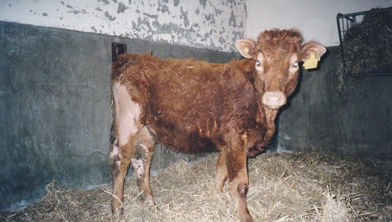
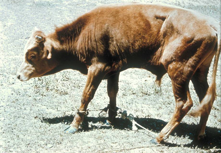
- Retarded growth
- Rough skin coat
- Loss of body weight
- Intermittent diarrhoea
- Emaciation
PM Findings:
- Hyperemic, erosive, ulcerative and necrotic lesion in oral mucosa, esophagus, rumen, reticulum, omasum and intestine.
- Destruction of Payer’s patches.
- Destruction of lymph nodes
- Colitis and proctitis characterized by hemorrhagic, ulcerative and necrotic changes.
- Histopathological changes in glandular epithelium of intestine: lesion consist of focal areas of ballooning of cells of stratum spinosum accompanied with nuclear keratolysis or pyknosis.

Diagnosis:
- On the basis of clinical findings
- On the basis of PM findings
- Isolation and identification of virus through conjunctival swab, sample of blood and feces.
- Serological test: CFT, ELISA, VNT, Immunofluorescence test
- Hematology: leukopenia, thrombocytopenia and neutropenia
- Biochemical test: Elevated BUN, hyponatremia, hypokalemia, hypochloremia
Differential Diagnosis:
- Rinderpest (RD):
- Heavy mortality, highly contagious
- Necrotic lesion on stratum spinosum
- Shooting diarrhoea
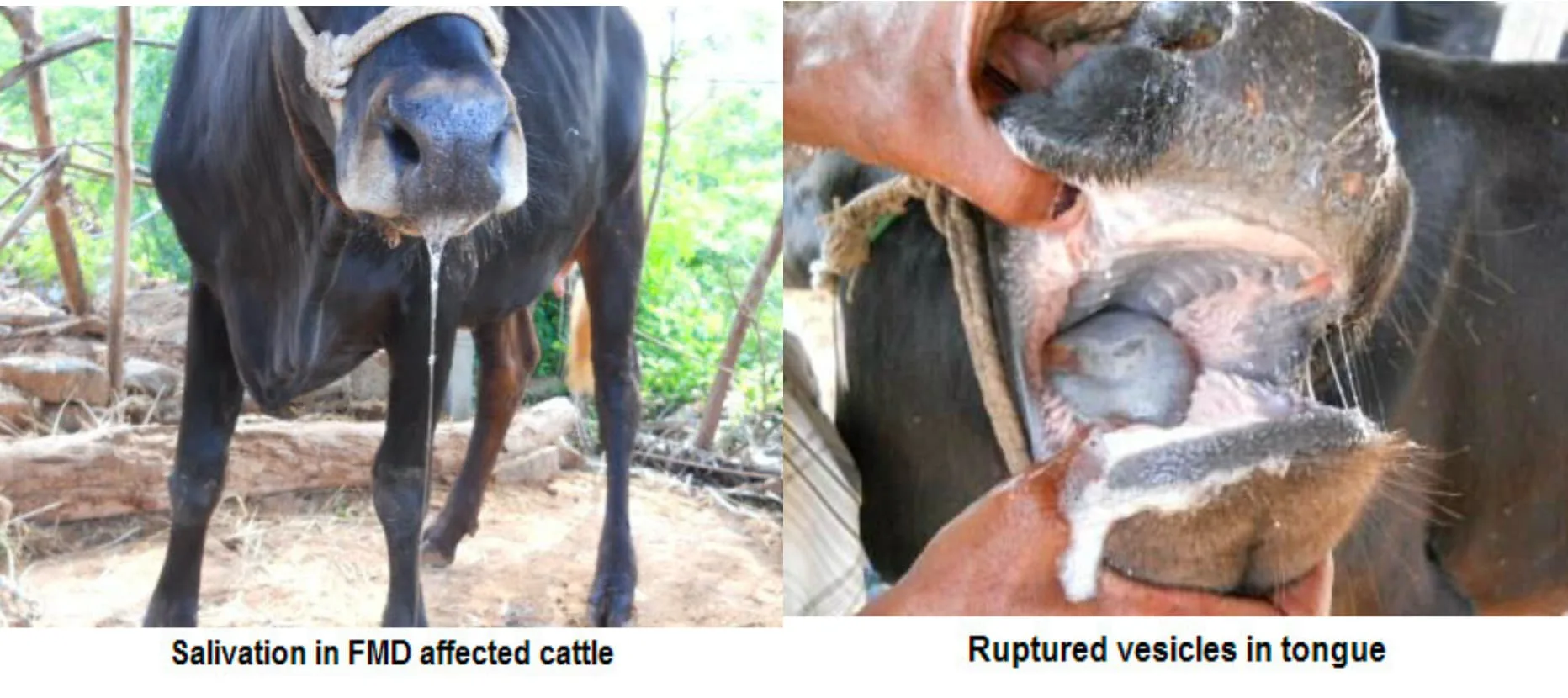
- FMD:
- Large and numerous vesicles in foot and mouth
- Stringy salivation
- Malignant Catarrhal Fever (MCF):
- Involvement of eyes
- High persistent fever
- Involvement of respiratory system
- Nervous manifestation (Encephalitis)

- Johne’s Disease:
- It is bacterial disease
- Chronic wasting disease
- Corrugation of intestine
- Demonstration of acid-fast bacilli by rectal scrapings.

Treatment and Control Measures:
- Since it is viral disease, it has no specific treatment. Symptomatic treatment is done.
- Antidiarrheal drugs are administered to prevent diarrhoea. Activated charcoal @ 1-2g/kg, PO repeated at 4-6 hour. Kaolin-pectin @3-4mL/kg, PO, 6-8h in horse.
- In cattle, most commonly used drugs are sulphonamides. Cotrimoxazole @15-20 mg/kg, b.wt. PO, OD
- Antibiotics are administered to prevent secondary bacterial infection.
- Fluid therapy to counter dehydration and ion loss in animals. D5, D10, RL can be administered to replenish electrolytes as well as water in body.
- Supplementation of animals with vitamins, minerals for body support.
- Infected calves should be culled to prevent spread of BVD.
- Vaccination in endemic areas:
- Modified live virus vaccine for 6 months calves.
- Inactivated vaccine; safe for pregnant cattle
- 2 doses- female before breeding, immunity is temporary.