Fowl Pox:
- It is slow spreading viral disease characterized by skin lesions.
- It was once widely prevalent worldwide, but with arrival of vaccination, the incidence has been greatly reduced.
Etiology:
- Avipox virus of Poxviridae family
- Enveloped, ds DNA virus
- It is largest virus known.
- It affects birds of both sexes, and all ages and breeds.
Epidemiology:
- Incubation period for fowl pox in chicken and turkey is typically 4-10 days.
- Cutaneous infection has low or moderate mortalities.
- Mortalities rate is higher in diphtheritic form.
- Disease is distributed globally.
- Virus mainly infects chicken and turkeys and has been reported to infect ducks, geese, pheasants, quail.
Transmission:
- Virus is usually transmitted by direct contact through abrasion of skin.
- Skin lesions (scabs) are source of aerosol exposure.
- Mosquitoes and other biting insect may serve as mechanical vectors.
- Virus can survive in dried scabs for months or even years.
Pathogenesis:
- Virus enters skin cell through abrasion/wound
- Virus then spread from cell to cell locally
- Some virus enters blood stream and causes viremia.
- Virus then reaches to internal organs like liver and spleen and secondary viremia occurs.
- Virus again enter into skin cells and a generalized disease can occur, but it is rare.
Clinical Findings:
- Disease appear in 2 forms:
- Cutaneous or skin form (dry pox)
- Diptheritic form (wet pox)
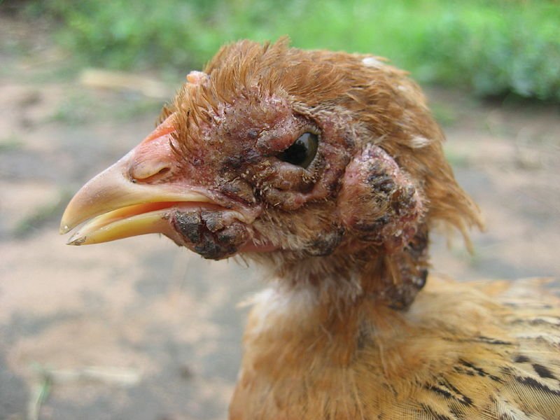
- Dry pox (Cutaneous or skin form):
- Pock lesion are seen on unfeathered skin; head, neck, legs and feet.
- First lesion appears as papule which progresses to vesicle to pustule and finally to crust or scab stage.
- After about 2 weeks, scab falls and healed area is left.

- Diptheritic form (wet pox):
- Small whitish nodules are observed in upper respiratory tract and digestive tract.
- These nodules merge together to form raised yellow cheesy plaques.
- Most lesion are found in mouth, larynx, trachea and esophagus.
- Difficulty in breathing and/or loss of appetite.
- Lesion in nose leads to nasal discharge and lesion in conjunctiva leads to ocular discharge. In rare cases, blindness occurs.
- Weakness, poor weight gain
- Egg production stops temporarily
Diagnosis:
- Based on history and slow spread of disease.
- Based on clinical findings and lesion.
- Histopathological examination of tissue stained with H&E reveals eosinophilic cytoplasmic inclusion bodies (Bollinger bodies). It is most commonly used method in diagnostic laboratory.
- FAT, Immunohistochemical method
- Elementary bodies (Borrell bodies) in inclusion bodies (Bollinger bodies) can be detected by light microscopy in smear from lesions.
- Isolation of virus in chorioallantoic membrane of developing chicken embryo.
- PCR
Treatment:
- No specific treatment available for disease.
- Symptomatic treatment is done. Wound/lesion are scratched, cleaned with antiseptics.
- Topical ointment for healing skin lesion is applied.
Control Measures:
- Vaccination of poultry flocks with live embryo or cell-culture propagated virus vaccine. It is done at first few weeks after hatching. Booster dose at 12-16 weeks is sufficient.
- Strict biosecurity measures such as sanitation and disinfection should be followed.
- Poultry houses, litter, equipments are sterilized and disinfected.
- Vector such as mosquitoes or biting insects should be controlled.
- Water ditches around poultry houses should be buried to control breeding of mosquitoes.