Pox Disease of Livestock
Cow pox:
- It is a mild contagious skin disease of cattle usually affecting the udder
- It is characterized by eruptions of pustular rash
- When virus transmitted to human via vaccination, it gives immunity against small pox.
Etiology:
- Vaccinia virus of genus orthopox virus
- Family: Poxviridae
- Virus produce hemorrhagic pock lesion on chorio-allantoic membrane of developing chick embryo
Transmission:
- Spread through direct contact with infected udders on cow’s teat
- Through direct contact to infected animals
- Rodent to human transmission
- Via hand of milkers
Pathogenesis:
- Virus enters through direct contact
- First replication occurs in skin or lungs
- Spread to regional LN followed by primary viremia
- Skin lesion appears as erythema and then macule
- Vesicles formation which becomes pustules. Pustules rupture to form scar.
Clinical Findings:
- Temperature is mild in nature
- Anorexia, suspended rumination and depression
- Udder is swollen, hot and sensitive
- Typical pock lesion appears on teat and udder
- Lesion on face of calf
- Swollen lymph nodes, fever
- Vesicles rupture leaving raw ulcerative areas that form scar
Diagnosis:
- Based on clinical signs, characteristic skin lesion
- Isolation of virus
Differential Diagnosis:
- FMD:
- Lesion in foot and mouth along with udder
.jpg)
- Pseudo cowpox:
- Scab formation in annular pattern
- Milker’s nodule formation on hand of milker

- Udder impetigo:
- It is caused by Staphylococcus aureus
- Lesion are discrete, superficial, thin walled and vesicle surrounded by zone of erythema.

Treatment:
- No specific treatment available. Symptomatic treatment is done
- NSAIDs; Meloxicam @0.2 mg/kg, b.wt. IM X OD for 3-5 days
- Broad-spectrum antibiotics; Streptopenicillin
- Application of povidone iodine; himax, salicyclic acid, benzoic acid ointment
Control Measures:
- Strict hygienic measures should be adopted.
- Isolation and segregation of affected cattle
- Disinfection of milking machine
- Hands of milker should be cleaved and washed with antiseptic soap before milking.
- Teat dipping into KMnO4 solution prior to dipping.
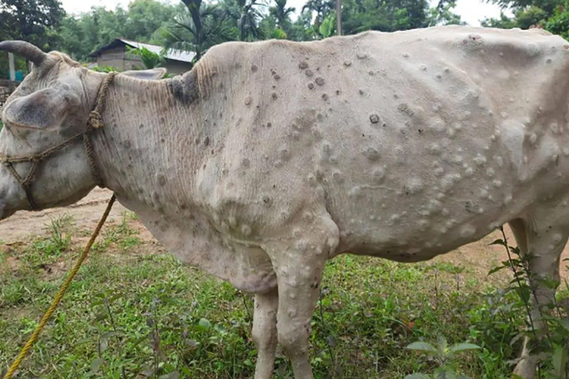
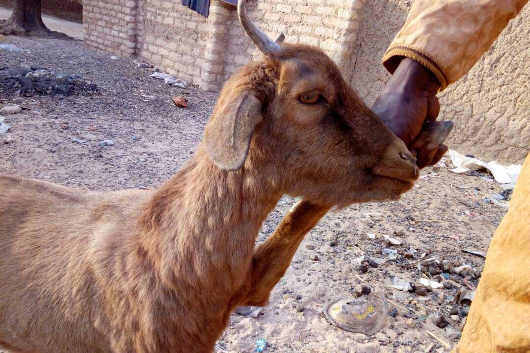
Goat pox:
- It is a malignant disease of goat characterized by fever and appearance of pock lesion.
- All breeds of goat irrespective of age and sex are susceptible.
Etiology:
- Capripox virus
Transmission:
- Through direct contact with affected animals
- Through wound and abrasions
- Virus present in skin papules are transmitted when infected animals rub their body on healthy animals
- Inoculation by biting insect
Pathogenesis:
- Inhalation of virus
- Virus reaches blood circulation via lymphatic duct causing primary viremia
- Virus then localizes to skin and target organs; lungs
- In lungs, lesion produce leading to bronchopneumonia
Clinical Signs:
- Incubation period is of 2-5 days
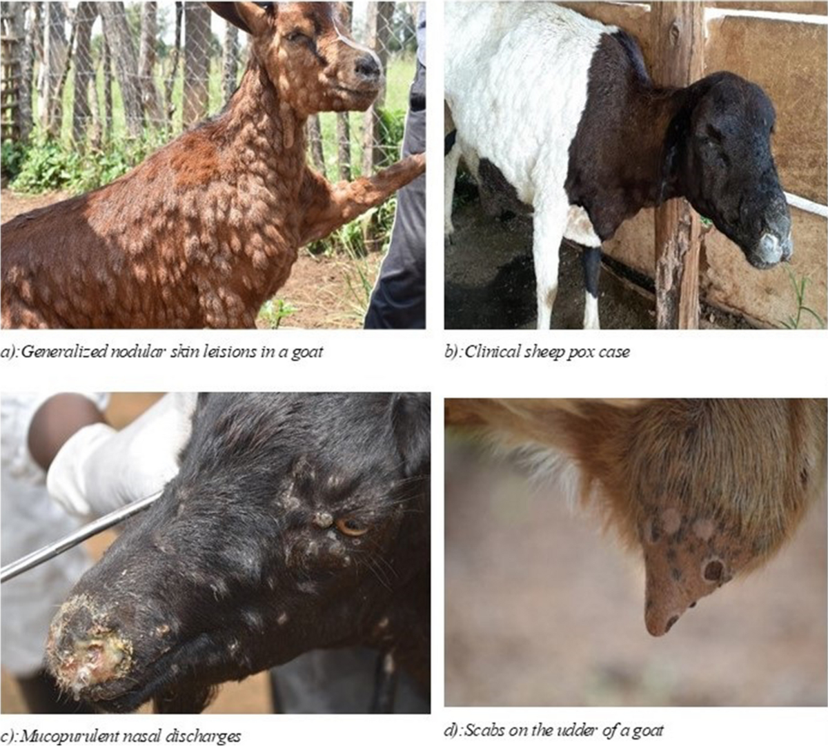
- Papules are preceded by red macules which is easily seen on white skin. First appear on hairless parts of skin
- Rhinitis, conjunctivitis
- Swollen superficial lymph nodes
- Eyelids are swollen and may completely cover the eyeball
- Mucopurulent ocular and nasal discharge when papules on conjunctiva and external nares become ulcerated.
- Mucus membrane of eyes, nose, lips, vulva and prepuce becomes necrotic
- Ultimately animals die due to laboured breathing as a result of broncho-pneumonia.
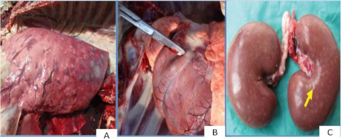
PM Findings:
- Pock lesion are seen in all parts of body, eg; lips, cheeks, snout, nostril, face, ear, feet, thigh, abdomen, eyelid, neck, teat, udder
- Papules are usually raised with dark brown centres and changes into vesicle containing cream-colored fluids within 2 days
- Hemorrhage in epidermis and edema of subcutis
- Ballooning degeneration of epithelial cells.
- Coagulative necrosis with pyknotic nuclei and infiltration of neutrophils
- Lung parenchyma shows focal areas of pneumonic changes characterized by congestion, red hepatization and exudation.

Diagnosis:
- On basis of clinical signs and symptoms
- Isolation of virus
- Serum neutralization test
- Gel diffusion test
- FAT
Differential Diagnosis:
- Contagious ecthyma:
- Lesion are usually in lips
- FMD
- Lesion mostly confined to mouth and feet.
Treatment and Control:
- Antiseptic or antibiotic ointment are applied.
- Scabs should be painted with povidone-iodine preparation
- Infected animals should be segregated.
- Infected milk shouldnot be fed to kids.
Sheep pox:
- Most serious pox disease of animals characterized by acute febrile condition and generalized development of pock lesion.

Etiology:
- Capri pox virus; DNA virus
- Relatively resistant to environmental temperature and stress
Susceptible Host:
- Sheep are naturally susceptible
- Younger sheep are more susceptible over older ones
- Merino breeds are more prone to disease than indigenous breeds.
Transmission:
- Direct contact with infected animals
- Droplet infection through nasal inhalation
- Through wound and abrasion
Pathogenesis:
- After virus gains entry, there is multiplication of virus with resultant viremia.
- Virus then distributed throughout body system
- Generalized skin lesion appears which turns into vesicles. Vesicles then turn into scab.
Clinical Signs:
Disease appears in 3 clinical forms:
i. Malignant form
ii. Mild (benign) form
iii. Abortive form
- Malignant form:
- Most common form of disease
- Affected sheep are dull and depressed with high fever; 106-107°F
- Pock lesion on eyelid, lips, nostrils, ears, cheeks, side of foreleg, thigh, scrotum, prepuce, vulva, under tail, chest region and buccal mucosa.
- Lesion begins as macules, then turn into papules which transform into large vesicles. Necrotic changes in the vesicle leads to scab formation.
- Mild form:
- Eruptions are confined around eye, lips and nose
- Usually noticed in adult sheep
- Abortive form:
- Ewe may abort and foetus shows pock lesion
- Lactating ewes shows the signs of mastitis due to lesion in udder
PM Findings:
- Characteristic papules, vesicles, pustules, and scabs are noted on cutaneous surface.
- Lesion observed in mucosa of respiratory and alimentary tract especially on trachea.
Diagnosis:
- On the basis of clinical signs and lesion
- Animal inoculation test: lymph of suspected material of affected sheep is inoculated into scarified surface of skin of suspected animal. Inoculated sheep shows high rise of temperature, papules and vesicle formation.
- CFT, Immunodiffusion test
- Gel diffusion test
Treatment and Control:
- Antiseptic or antibiotic ointment are applied.
- Scabs should be painted with povidone-iodine preparation
- Infected animals should be segregated.
- Infected milk shouldnot be fed to lambs
- Vaccination; attenuated or killed vaccine, freeze dried vaccine is administered.
Swine pox:
Syn: Contagious impetigo, Louse borne dermatitis
- It is highly contagious viral disease of pigs characterized by skin eruptions and cutaneous lesion.
- It is mostly seen in young pigs 3-6 weeks old but all age is affected.
Etiology:
- Suipox virus
- Virus can be grown in tissue culture.
Epidemiology:
- Disease is distributed world-wide.
- It is more frequently seen on areas where intensive breeding program has been undertaken.
- It has been recorded as an outbreak in large white pig from Andhra Pradesh, India.
- Cases have been reported in wild boars, suggesting a potential wildlife reservoir for the virus.
- The severity of the disease can vary, with younger pigs experiencing more noticeable symptoms and potentially higher mortality rates.
Transmission:
- Through direct contact with infected pig
- Rubbing of infected skin on healthy pig
- Through vectors; biting lice; Hematopinus suis
Pathogenesis:
- Virus enters through skin abrasion or bites
- Replication of virus in epidermal cells
- Formation of vesicles
- Vesicles ruptures and turns into scab which heal or drop off
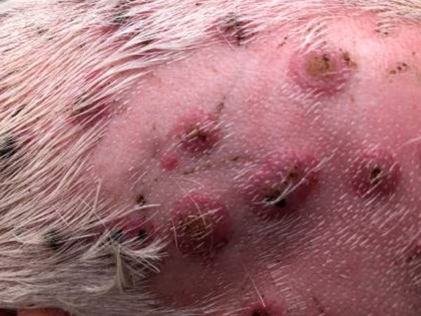
Clinical Findings:
- Incubation period; 1 week
- Affected pigs are dull and depressed with dejected appearance.
- High fever; 105-107°F with nasal and ocular discharge
- Pock lesion appears over snouts, eyelids, inner aspect of thigh, abdomen
- Lesion are vesicular at first which turns to papules. Red brown scab develops on skin within 8-11 days.
PM Findings:
- Multifocal, popular to pustular lesion are seen on ventral abdomen, inner thighs, and perioral/ocular region.
- Ballooning degeneration of epidermal cells.
- Fluid accumulation within abdominal cavity
- Eosinophilic inclusion bodies are found within cytoplasm of keratinocytes.

Diagnosis:
- Based on clinical findings
- Based on PM findings
- Isolation and identification of virus
Differential Diagnosis:
- Mites infestation
- Scabies; Itching is cardinal sign
- Ringworm: Lesion are crusty in nature
Treatment:
- There is no specific treatment for disease.
- It is better to use Butox @3-5 ml/litre of water
- Ivermectin @1ml/50 kg, b.wt., SC, SD
Control Measures:
- Vaccination is not attempted.
- Recovery from infection renders the pig immune to disease.
- Lice should be controlled in pigs. Acaricides like lindane, trichlorophan, amitraz, etc may be applied.
- Animals should be cleaned and disinfected to keep the premises louse free.
- Isolation of affected animals from herd.
- Regular inspection of skin of newly purchased pigs for signs of pox lesions.